


代理商
江苏艾洛特生物科技有限公司
入驻年限:13 年
- 联系人:
陆小姐
- 所在地区:
江苏 南京市 江宁区
- 业务范围:
试剂、技术服务、细胞库 / 细胞培养、ELISA 试剂盒
- 经营模式:
生产厂商 代理商
推荐产品


公司新闻/正文
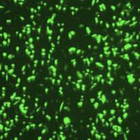
角质化细胞
人阅读 发布时间:2014-06-19 16:42
角质化细胞
“正常人表皮角质形成细胞(NHEK)少年包皮,单一捐献者” ,(Normal Human Epidermal Keratinocytes (NHEK) juvenile foreskin, single donor,500,000 cryopreserved cells)。
以下是产品的详细信息,可点击链接查看:
http://www.promocell.com/fileadmin/promocell/PDF/C-12001.pdf
http://www.promocell.com/fileadmin/promocell/MSDS/C-12001.pdf
正常的人类皮肤角质形成细胞(NHEK) 分离自青少年包皮的表皮,以及成年人各个部位的皮肤,例如面部、乳房、腹部和大腿*。这些细胞是表皮中的主要细胞类型,占据了细胞总数的90%。
表皮角质化细胞来自皮肤基底层,能穿过表皮各个层次上移。在这个移动过程中,这些细胞逐渐分化,并产生形态学改变,直到到达皮肤的角质层,在角质层,这些细胞形成一层无核的、扁平的和高度角质化的鳞状细胞。这层细胞形成一层有效的屏障,防止外来物质进入到机体,同时尽可能降低水分流失。
角质化细胞也能够产生一系列细胞因子、生长因子、白细胞介素和补体因子。因此,角质化细胞对伤口愈合、炎症和免疫应答都具有重要作用。
The cells have been tested: Cytokeratin positive
http://www.promocell.com/fileadmin/promocell/Kapitelbilder/Keratinocytes_2.jpg
应用范围
- 伤口愈合研究
- 皮肤发育和分化研究
- 皮肤病学研究
- 药物摄取研究
- 制药学测试
- 化妆品和毒理学检测
- 肿瘤学
- 细胞角蛋白阳性
- 青少年包皮或成年人不同部位的皮肤上来源的也有供应
- 单一捐献者和混合捐献者来源的都有供应
推荐的培养基及试剂
相关产品
- NHEK.f 细胞团块
- NHEK.f 混合来源的细胞团块
- NHEK 成年人来源的细胞团块
- NHEK 混合成年人来源的细胞团块
- 正常人类真皮成纤维细胞
- 正常人类表皮黑色素细胞
- PromoFectin 转染试剂
以下是NHEK细胞的相关问题,可参考:
http://www.promocell.com/index.php?id=1049&tx_na15knowledgebase_pi1[categories][]=3&no_cache=1&tx_na15knowledgebase_pi1[sword]=NHEK
What's the difference between NHEK.f (C-12001) and NHEK.f pooled (C-12005)?
NHEK.f are isolated from tissue samples (foreskin) of single donors, aged between 1-10 years.
NHEK.f pooled are prepared from the foreskins of 3 individual donors. The cells of each donor are expanded in separate TC vessels and the cells are pooled after secondary culture, before cryopreservation.
Both NHEK from single donor and NHEK pooled are in P2 after thawing.
Do the PromoCell keratinocytes need feeder cells?
If you are growing our NHEK in PromoCell Keratinocyte Growth Medium 2 (C-20011), you don't need any feeder cells. The cells will grow as a monolayer in conventional tissue culture flasks.
How does PromoCell determine the phototype of skin tissue donors?
We use the classification of skin types (phototype I-VI) according to Fitzpatrick. This is determined by the patient's skin colour (white, brown or black skin), color of eyes and hair, and by the burning/tanning ability, i.e. by the amount of melanin pigment in the skin.
I: Pale white skin, blue/hazel eyes, blond/red hair, always burns, does not tan
II: Fair skin, blue eyes, burns easily, tans poorly
III: Darker white skin, tans after initial burn
IV: Light brown skin, burns minimally, tans easily
V: Brown skin, rarely burns, tans darkly easily
VI: Dark brown or black skin, never burns, always tans darkly
Donors with phototype I and II are classified as L (lightly pigmented), with phototype III and IV as M (moderately pigmented), and with phototype V and VI as D (darkly pigmented).
Information on the phototype is available for most our cell lots isolated from juvenile or adult skin.
Is it possible to obtain other cell types, e.g. keratinocytes, from the same fibroblast donor?
Yes, in principle it is possible to isolate keratinocytes (NHEK), melanocytes (NHEM), dermal microvascular endothelial cells (HDMEC), and fibroblasts (NHDF) from the same skin sample.
As we have a strict quality control system, some cell preparations can fail to pass QC and aren't released for sale. Therefore, please contact our Technical Customer Service if you need different cell types from the same donor so that we can check our inventory.
What is the trypsinization time with PromoCell keratinocytes?
The trypsinization time of primary keratinocytes (and epithelial cells in general) is usually longer than of other cells. Depending on the cell lot, it takes between 5 and 10 min at room temperature. You can accelerate the detachment by gently tapping the flask as soon as the cells shrink and round up. Alternatively, you can use accutase (C-41310) at 37°C which has been shown to not affect cellular viability even at longer incubation times.
参考文献 ——以下是近年来使用我们PromoCell的NHEK细胞进行试验研究,在国际知名杂志所发表的文章,如下:
Residual antimicrobial effect of chlorhexidine digluconate and octenidine dihydrochloride on reconstructed human epidermis
Mueller et al.; Skin Pharmacol Physiol. 2014;27(1):1-8
Demonstration of a melanoma-specific CD44 alternative splicing pattern that remains qualitatively stable, but shows quantitative changes during tumour progression
Raso-Barnett et al.; PLoS One. 2013;8(1):e53883
Aryl hydrocarbon receptor repressor (AhRR) function revisited: repression of CYP1 activity in human skin fibroblasts is not related to AhRR expression
Tigges et al.; J Invest Dermatol. 2013 Jan;133(1):87-96
Plant-derived human collagen scaffolds for skin tissue engineering
Willard et al.; Tissue Eng Part A. 2013 Jul;19(13-14):1507-18
Lipopeptide biosurfactant pseudofactin II induced apoptosis of melanoma A 375 cells by specific interaction with the plasma membrane
Janek et al.; PLoS ONE. 2013;8(3):e57991
Infliximab induces downregulation of the IL-12/IL-23 axis in 6-sulfo-LacNac (slan)1 dendritic cells and macrophages
Brunner et al.; J Allergy Clin Immunol. 2013 Nov;132(5):1184-1193.e8
Heparin increases the infectivity of human Papillomavirus type 16 independent of cell surface proteoglycans and induces L1 epitope exposure
Cerqueira et al.; Cell Microbiol. 2013 Nov;15(11):1818-36
Follicular dermal papilla structures by organization of epithelial and mesenchymal cells in interfacial polyelectrolyte complex fibers
Lim et al.; Biomaterials. 2013 Sep;34(29):7064-72
Amygdalin analogues inhibit IFN-gamma signalling and reduce the inflammatory response in human epidermal keratinocytes
Paoletti et al.; Inflammation. 2013 Dec;36(6):1316-26
A purified Feverfew extract protects from oxidative damage by inducing DNA repair in skin cells via a PI3-kinase-dependent Nrf2/ARE pathway
Rodriguez et al.; J Dermatol Sci. 2013 Dec;72(3):304-10
The transcription factors TBX2 and TBX3 interact with human Papillomavirus 16 (HPV16) L2 and repress the long control region of HPVs
Schneider et al.; J Virol. 2013 Apr;87(8):4461-74
Vinblastine-induced apoptosis of melanoma cells is mediated by Ras homologous A protein (Rho A) via mitochondrial and non-mitochondrial-dependent mechanisms
Selimovic et al.; Apoptosis. 2013 Aug;18(8):980-97
IL-17A and IFN-gamma synergistically induce RNase 7 expression via STAT3 in primary keratinocytes
Simanski et al.; PLoS One. 2013;8(3):e59531
Retargeting of rat parvovirus H-1PV to cancer cells through genetic engineering of the viral capsid
Allaume et al.; J Virol. 2012 Apr;86(7):3452-65
Resistance to HSV-1 infection in the epithelium resides with the novel innate sensor, IFI-16
Conrady et al.; Mucosal Immunol. 2012 Mar;5(2):173-83
Xenobiotic metabolism capacities of human skin in comparison to a 3D-epidermis model and keratinocyte-based cell culture as in vitro alternatives for chemical testing: phase II enzymes
Götz et al.; Exp Dermatol. 2012 May;21(5):364-9
Human keratinocytes' response to injury upregulates CCL20 and other genes linking innate and adaptive immunity
Kennedy-Crispin et al.; J Invest Dermatol. 2012 Jan;132(1):105-13
Combination of sulindac and dichloroacetate kills cancer cells via oxidative damage
Ayyanathan et al.; PLoS One. 2012;7(7):e39949
High thrombin concentrations in fibrin sealants induce apoptosis in human keratinocytes
Gugerell et al.; J Biomed Mater Res A. 2012 May;100(5):1239-47
A subunit of eukaryotic translation initiation factor 2 alpha-phosphatase (CreP/PPP1R15B), regulates membrane traffic
Kloft et al.; J Biol Chem. 2012 Oct 12;287(42):35299-317
Estradiol protects the dermal hyaluronan/versican matrix during photoaging by release of epidermal growth factor from keratinocytes
Rock et al.; J Biol Chem. 2012 Jun 8;287(24):20056-69
Keratinocyte growth factor induces gene expression signature associated with suppression of malignant phenotype of cutaneous squamous carcinoma cells
Toriseva et al.; PLoS One. 2012;7(3):e33041
Metal allergens nickel and cobalt facilitate TLR4 homodimerization independently of MD2
Raghavan et al.; EMBO Rep. 2012 Nov 30;13(12):1109-15
Centrosomal localization of the Psoriasis candidate gene product, CCHCR1, supports a role in cytoskeletal organization
Tervaniemi et al.; PLoS One. 2012;7(11):e49920
Antialarmin effect of tick saliva during the transmission of Lyme disease
Marchal et al.; Infect Immun. 2011 Feb;79(2):774-85
Decisive role of tumor necrosis factor-α for spongiosis formation in acute eczematous dermatitis
Kerstan et al.; Arch Dermatol Res. 2011 Nov;303(9):651-8
Activation and differentiation of mesenchymal stem cells
Mishra and Banerjee; Methods Mol Biol. 2011;717:245-53
Differential methylation of the HPV 16 upstream regulatory region during epithelial differentiation and neoplastic transformation
Vinokurova and von Knebel Doeberitz; PLoS One. 2011;6(9):e24451
A new amyloidosis caused by fibrillar aggregates of mutated corneodesmosin
Caubet et al.; FASEB J. 2010 Sep;24(9):3416-26
Nicotinic acid- and monomethyl fumarate-induced flushing involves GPR109A expressed by keratinocytes and COX-2-dependent prostanoid formation in mice
Hanson et al.; J Clin Invest. 2010 Aug 2;120(8):2910-9
询价列表

暂时没有已询价产品

快捷询价 发送名片
当你希望让更多商家联系你时,可以勾选后发送询价,平台会将你的询价消息推荐给更多商家。